Destruction/ Repair/ Remodelling Phases
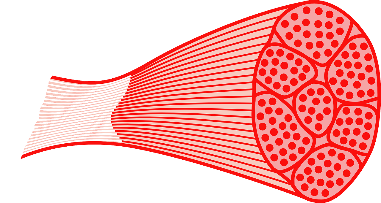
Skeletal muscle ranges from 40 to 50% in men of total body weight and 25 to 35% in women and can reach to 65% in weightlifters.
Muscle fibres can be divided into two types with 50% composed of slow twitch/ contraction fibres (type I) and 50% of fast twitch contraction fibres (type II). Postural muscles have a larger quantity of type I fibres with presenting to be a slow contraction speed but a greater resistance to fatigue.
Type II fibers fast contraction fibres) can be subdivided into types IIA and IIB. Type IIA fibres are known as intermediate layers that have larger quantities of mitochondria and myoglobin than seen in type IIB fibres and are thus more resistant to fatigue. Type II fibers (fast twitch fibres) can be transformed into type I fibers (slow twitch fibres) through training.
According Barroso and Thiele (2015), the largest portion of muscle injuries occur during sports activities and correspond to 10 to 55% of all sporting injuries.
The Physiology of Injuries and The Three Stages of Muscle Repair Post Injury
1. Destruction/ Inflammation Phase
1. Destruction/ inflammation Phase: Muscle degeneration and inflammation occurs throughout the first few days post injury and is characterised via the rupture and necrosis of muscle fibres. Many pro-inflammatory molecules such as cytokines, chemokine and growth factors are secreted by neutrophils.
Macrophages are a type of immune cell that are involved during the inflammation stage that are defined as pro-inflammatory macrophages. These cells act during the first few days post injury and contribute towards cell lysis or the 'eat' and 'clean away' the removal of cellular debris and stimulate myoblast proliferation. M2 macrophages, defined as anti-inflammatory macrophages, act two to four days post injury and help with muscle repair by promoting myotubule formation (2).
Treatment Implications: During this phase, immobilisation with braces, splints, crutches or taping technique application might be appropriate. Brief immobilisation for 3-7 days can assist with minimising the pain and swelling within the injured tissue. Application of RICE (Rest, ice, compression, elevation) can help reduce the inflammation and promote wound healing.
2. Repair/ Regeneration Phase
2. Repair/ Regeneration/ Remodelling Phase: During this stage, damages muscle undergoes repair and reorganisation to restore the muscle's structure and function.
Macrophage is introduced into the injured site. A macrophage “eats” and will “cleans away” dead tissue and dry blood caused by injury. Once this is complete, another cell called a satellite cell is released into the injured area. Satellite cells transform into myoblast cells, which group together to create new muscle fibres. Fibroblasts also produce connective tissue at the injured site and it is a combination of connective tissue and muscle fibres that repair the injured muscle. In addition, new blood vessels (angiogenesis) and nerves generate during this phase. This repair phase commonly peaks about two weeks after injury (2).
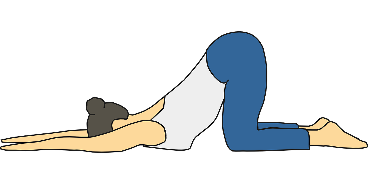
Treatment Implications: Moving and mobilising muscles can encourage faster regrowth of blood vessels and muscle fibres which will reduce scarring formation and increase tensile strength of muscle fibres. The expertise of physical therapists is important at this phase, as they can help select appropriate rehabilitation exercises. Initially, gentle exercises called isometrics exercises might be prescribed whereby the muscle contracts but there is no movement of a joint or limb. Next, progression to isotonic exercises would be prescribed to strengthen the muscle through its full range of motion. This, in combination with gentle stretching, is standard during the repair phase.
A few soft tissue massage techniques can be viewed below:
3. Remodelling Phase
This stage can overlap with the repair phase, the regenerating of muscle fibers and connective tissue continue to mature and are being oriented into the final scar tissue. This stage is important for the manner in which the scar tissue is being orientated. Typically, muscle tissue is orientated in straight lines. When the tissue repairs itself, the mixture of new muscle fibres and connective tissue is randomly orientated. Hands-on physical therapy treatment during this phase can assist the new tissue to regenerate into parallel lines, like a pile of straws, instead of a big knotted lumps, like a piece of cord or muscle 'knot', which is sensitive and painful.
Treatment implications: Instrument assisted soft tissue massage techniques and specialised massage tools may be used by physical therapists along with medical acupuncture to help reduce the tension within muscles and muscular knots. This is important for reducing the likelihood of reinjuring the tissue. Returning to regular functional activity by normalising joint mechanics, regaining full strength through isokinetic exercises and sports-specific training can occur during this time.
References
1. Barroso, G. C., Thiele, E. S. (2011) Muscle Injuries in Athletes, Rev Bras Ortop., 46; 4: 354-58.
2. Koh, T. J., Dipietro, L. A. (2011) Inflammation and Wound Healing: The Role of the Macrophage, NIH, 13, e23: 1-14.
